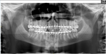
The emergency room evaluation reveals a patient in acute distress from an injury to her chest and jaw. During the MVA she hit her chin on the right side; she has bruising and localized swelling on the right side but she cannot get her teeth together on the left and she is having pain in the left preauricular area. A pantograph and CT scan show fractures in the neck of the left condyle and body of the right mandible.
She is scheduled for a OMFS consult for the management of the fracture.
Think About the Following Questions
- What level of discomfort should the patient expect to have from her chest injury and jaw fracture?1
- What imaging is appropriate for assessing a fracture of the mandible?2
- What complications might one expect after a fracture of the mandible?3, 4
References
- Throckmorton GS, Ellis E 3rd. Recovery of mandibular motion after closed and open treatment of unilateral mandibularcondylar process fractures. Int J Oral Maxillofac Surg. 2000 Dec;29(6):421-7. PMID:11202321
- Dreizin D, Nam AJ, Tirada N, Levin MD, Stein DM, Bodanapally UK, Mirvis SE, Munera F. Multidetector CT of Mandibular Fractures, Reductions, and Complication: A Clinically Relevant Primer for the Radiologist. Radiographics. 2016 Sep-Oct;36(5):1539-64. doi: 10.1148/rg.2016150218. Review. PMID:27618328
- Meara DJ, Jones LC. Controversies in Maxillofacial Trauma. Oral Maxillofac Surg Clin North Am. 2017 Nov;29(4):391-399. doi: 10.1016/j.coms.2017.06.002. Review. PMID:28987223
- García-Guerrero I, Ramírez JM, Gómez de Diego R, Martínez-González JM, Poblador MS, Lancho JL. Complications in the treatment of mandibular condylar fracture: Surgical versus conservative treatment. Ann Anat. 2018 Mar;216:60-68. doi: 10.1016/j.aanat.2017.10.007. Epub 2017 Dec 6. Review. PMID:29223659
OMFS Consult Findings
- The patient’s jaw range-of-motion (ROM) shows a somewhat limited vertical ROM of 35 mm with a deflection to the left upon opening. (She has pain with any vertical jaw movement making her pain-free ROM zero and her ROM with pain 35 mm). She can move laterally to the left 7 mm but she has pain with this movement in the left deep masseter area. She cannot move her jaw in a right lateral movement.
- The palpation examination shows severe pain to palpation just inferior to the left TMJ area. She is also sensitive to palpation over the right anterior mandible (the area where her jaw was hit).
- She has contact on her right posterior teeth as determined by shimstock but no contact on her left posterior teeth.
- The recommendation from the OMFS consult is for immediate reduction of the fracture due to its effect on the patient’s occlusion and eventual jaw function.
Think About the Following Questions
- What is the rationale for measuring pain-free vs pain-full jaw ROM?
- Why can the patient not move her jaw to the right?
- What criteria determine how a sub-condylar jaw fracture should be managed?
References
- Rozeboom A, Dubois L, Bos R, Spijker R, de Lange J. Open treatment of unilateral mandibular condyle fractures in adults: a systematic review. Int J Oral Maxillofac Surg. 2017 Oct;46(10):1257-1266. doi: 10.1016/j.ijom.2017.06.018. Epub 2017 Jul 18. Review. PMID:28732561
- Rozeboom AVJ, Dubois L, Bos RRM, Spijker R, de Lange J. Closed treatment of unilateral mandibular condyle fractures in adults: a systematic review. Int J Oral Maxillofac Surg. 2017 Apr;46(4):456-464. doi: 10.1016/j.ijom.2016.11.009. Epub 2016 Dec 7. PMID: 27955799
- Nitzan DW, Palla S. "Closed Reduction" Principles Can Manage Diverse Conditions of Temporomandibular Joint Vertical Height Loss: From Displaced Condylar Fractures to Idiopathic Condylar Resorption. J Oral Maxillofac Surg. 2017 Jun;75(6):1163.e1-1163.e20. doi: 10.1016/j.joms.2017.01.037. Epub 2017 Feb 7. PMID:28257719
OMFS Plan Summary
- The initial surgical plan called for the placement of arch bars on the maxilla and mandible and ligation of the arch bars to their respective arch and to the bar on the opposing arch.
- The patient was taken to the OR for oral surgery under general anesthesia via nasal intubation.
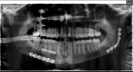
- In the OR, rigid fixation of the fractured neck of the left condyle and right mandible is provided by plating placed across the fractures and fixing the plates with screws on either side of the fracture site.
- The arch bars are then removed after adequate jaw function and a stable occlusion are determined.
Think About the Following Questions
- How does one determine that the occlusion is stable?
- What are your expectations for post-surgical rehabilitation of jaw function?
Reference
- Throckmorton GS, Ellis E 3rd . Recovery of mandibular motion after closed and open treatment of unilateral mandibular condylar process fractures. Int J Oral Maxillofac Surg. 2000 Dec;29(6):421-7. PMID:11202321
Oral Surgery: Management of Sub-Condyle Fracture
Ms. Tatum is a 30-year-old female who was involved in an MVA admission to the Hospital. She struck her lower jaw on the right on the automobile dashboard. Clinical and CT imaging demonstrated a fracture between the right mandibular first molar and second premolar as well as a laterally displaced sub condylar fracture on the left side. She was initially treated with closed reduction with maxillomandibular fixation under local anesthesia in the EW. Adequate reduction of the right mandibular body was obtained; however, persistent lateral dislocation of the proximal portion of the sub condylar fracture persisted.
The patient also expressed a desire to be removed from maxillomandibular fixation as soon as possible. The option for open reduction and internal fixation were discussed and it was agreed that the left sub condylar fracture would be approached via an endoscopic incision in the right submandibular region and the right mandibular body fracture would be approached by a right submandibular incision.
At surgery good reduction was obtained with rigid plates for both fractures, her occlusion was stable and reproducible and the maxillomandibular fixation was removed.
Panoramic Radiograph with the Maxillomandibular Fixation in Place
Fractures are marked with arrows.

Post-Operative Panoramic Radiograph
Shows rigid plates in place and the fractures reduced.


